

Moles and Melanoma: How to Examine Your Skin Using the ABCDE Rule
Melanoma detected at stage 1 has a 98% five-year survival rate. Detected at stage 4, that drops to 23%. The difference is finding it early.
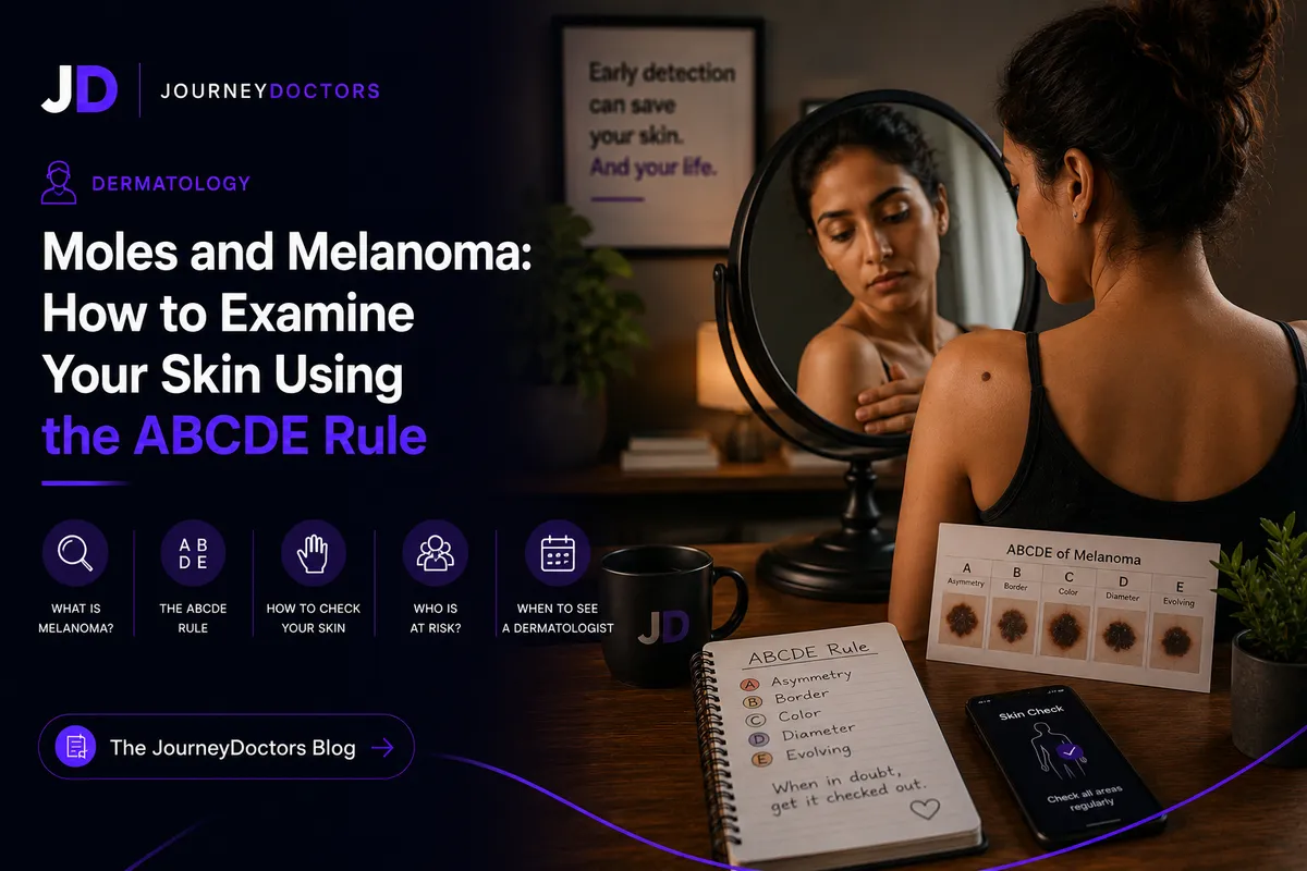
Melanoma is the most dangerous form of skin cancer, responsible for the majority of skin cancer deaths despite being far less common than basal cell and squamous cell carcinoma. As a dermatologist, I consider skin self-examination one of the highest-value preventive habits available to patients, precisely because the survival difference between early and late detection is so dramatic. Melanoma detected at stage 1, when it is confined to the top layer of skin and is thin, has a five-year survival rate of approximately 98 percent. By stage 4, when it has spread to distant organs, that figure drops to around 23 percent. The difference is detection before spread occurs, and regular skin examination by both patients and clinicians is the most reliable route to that early detection.
What Melanoma Is
Melanoma arises from melanocytes, the pigment-producing cells of the skin. It can develop in existing moles or in apparently normal skin. Its defining clinical danger is its tendency to metastasize: unlike basal cell carcinoma, which almost never spreads beyond the local site, melanoma can spread through the lymphatic system and bloodstream to lymph nodes, lungs, liver, brain, and bone. This capacity for distant spread is what makes early detection life-saving.
Ultraviolet radiation is the primary environmental risk factor. Both UVA and UVB rays damage DNA in melanocytes. Cumulative UV exposure and episodes of severe blistering sunburn, particularly in childhood, are the most clinically significant risk exposures. Indoor tanning beds, which emit concentrated UV radiation, significantly increase melanoma risk and are associated with a 59 percent increased risk when used before age 35 in multiple studies.
Risk Factors
Several factors increase melanoma risk beyond average. Fair skin, light hair, and light eyes reflect lower melanin content and reduced natural UV protection. A large number of moles (more than 50) or atypical moles (dysplastic nevi), which are moles with irregular features, significantly increase risk. A personal history of melanoma dramatically increases the risk of a second primary melanoma. A family history of melanoma, particularly in a first-degree relative, doubles or triples risk and suggests possible hereditary predisposition involving genes like CDKN2A or CDK4. Immunosuppression, whether from organ transplantation, HIV, or certain medications, increases melanoma risk. People who have had extensive cumulative sun exposure, particularly outdoor workers and people who grew up in high UV environments, have higher lifetime risk.
Importantly, melanoma also occurs in people without obvious risk factors, in skin tones across the full spectrum, and in body locations not typically exposed to the sun, including the soles of the feet, under the nails (subungual melanoma), and on mucosal surfaces. Acral lentiginous melanoma, which occurs on palms, soles, and under nails, disproportionately affects people with darker skin tones and is the most common subtype in Black and Asian populations. This matters because melanoma in people with darker skin is often diagnosed later due to the assumption that dark skin confers complete protection.
The ABCDE Rule
JourneyDoctors
Not sure if this applies to you?
Describe your symptoms to Dr. Maya — our AI GP — and get a real clinical response in under a minute. Free to start.
Talk to Dr. MayaThe ABCDE criteria provide a systematic framework for evaluating suspicious lesions. Learning and applying these criteria during monthly skin self-examination is the practical tool that enables early detection.
A: Asymmetry
A benign mole is typically symmetrical: if divided in half, both sides would appear roughly similar. Melanoma lesions are often asymmetrical: one half looks different from the other. Asymmetry alone is not diagnostic, but it is a reason to have the lesion evaluated by a dermatologist.
B: Border
Benign moles have smooth, well-defined borders. Melanoma often has irregular, ragged, notched, or blurred borders that make the edge difficult to define clearly. The ink spreading unevenly from a pen nib is a useful visual analogy for melanoma border irregularity.
C: Color
Benign moles are typically a uniform shade of brown. Melanoma often contains multiple colors within a single lesion: shades of brown, black, red, white, and blue. Color variation within a single mole is more clinically significant than absolute darkness. A dark mole that is uniformly dark is less suspicious than a smaller mole with multiple color variations.
D: Diameter
Melanoma lesions are often larger than 6 mm in diameter, approximately the size of a pencil eraser. This criterion is the weakest of the five because small melanomas exist and because many benign moles are larger than 6 mm. Diameter is most useful in combination with the other criteria. Any lesion growing in diameter, regardless of its current size, warrants evaluation.
E: Evolution
The E criterion is often the most clinically useful. Any mole or skin lesion that changes over time, in size, shape, color, or character, requires evaluation. Benign moles remain stable over years. Symptoms such as itching, bleeding, or crusting from a mole that previously caused no symptoms are also relevant changes that should prompt dermatological review. The evolution criterion captures the dynamic, growing character of melanoma that the static ABCD criteria cannot.
The Ugly Duckling Sign
An additional clinical heuristic is the "ugly duckling" sign: a lesion that looks distinctly different from all other moles on a given person's body warrants attention, even if it does not obviously fulfill the ABCDE criteria. Most people's moles share similar characteristics (a baseline signature); a lesion that stands out from this pattern is suspicious regardless of whether it is lighter or darker, larger or smaller than the average.
How to Examine Your Skin
Monthly skin self-examination should be systematic and performed in good lighting with a full-length mirror and a hand mirror for hard-to-see areas. Start with the face, scalp (use a comb to part the hair, or have a partner check), neck, and ears. Move to the chest, abdomen, and front of the arms and legs. Use the hand mirror to check the back, back of the arms and legs, buttocks, and soles of the feet. Check between the toes and under the nails. For people with many moles or high risk, body mapping with photographs helps track changes over time.
When to See a Dermatologist
Any lesion that meets ABCDE criteria, shows the ugly duckling sign, is new and growing rapidly, or symptoms from a previously stable mole should be evaluated by a dermatologist. The appropriate response to a suspicious lesion is excisional biopsy, not observation. Dermoscopy (examination with a dermatoscope, which provides magnified illuminated views of skin lesions) significantly improves the accuracy of clinical diagnosis and is standard in specialist dermatological practice.
For high-risk individuals, annual total body skin examination by a dermatologist is appropriate. This includes people with a personal or family history of melanoma, many atypical moles, or multiple significant risk factors. JourneyDoctors dermatologists can evaluate suspicious lesions and advise on appropriate follow-up. Consultations start at $19.
Disclaimer: This article is for educational purposes only and does not constitute medical advice. If you have a suspicious mole, please seek in-person evaluation by a qualified healthcare professional.
Get proper care
Ready to speak with a specialist?
If anything in this article sounds familiar, the right next step is a proper evaluation. JourneyDoctors connects you with a specialist in minutes. Consultations from $19.
See a specialist nowFrequently Asked Questions
Can melanoma develop in dark skin?
Yes. While melanoma is less common in people with darker skin tones, it does occur and is more likely to be diagnosed at a later stage in these populations, partly because of assumptions about risk and partly because acral lentiginous melanoma (on palms, soles, and under nails) is more common and may be overlooked in locations not typically examined. Skin self-examination should include these locations regardless of skin tone.
Is a dark mole always dangerous?
No. Many dark moles are benign, and the color criterion in ABCDE is about color variation within a lesion rather than absolute darkness. A uniformly dark, stable, symmetric mole with a smooth border is reassuring. A lesion with multiple color tones, irregular borders, and recent changes is more concerning, even if it is not particularly dark overall.
What happens if I have a suspicious mole biopsied?
A suspicious lesion is typically excised (removed with a small margin of normal skin) and sent for pathological examination. If the pathology confirms melanoma, additional surgery may be needed to clear wider margins, and sentinel lymph node biopsy may be recommended to assess spread. Treatment depends on stage: early stage melanoma is treated surgically with excellent outcomes; more advanced disease may require immunotherapy, targeted therapy, or other systemic treatment.
Does sunscreen prevent melanoma?
Yes, with strong evidence. Regular broad-spectrum sunscreen use reduces the risk of both squamous cell carcinoma and melanoma. A study in Queensland, Australia, randomized participants to daily sunscreen versus discretionary use and demonstrated a significant reduction in melanoma incidence and mortality in the daily sunscreen group over a 15-year follow-up. SPF 30 or higher, broad-spectrum (UVA and UVB protection), reapplied every two hours during sun exposure, is the evidence-based recommendation.
How often should I have a professional skin check?
For people at average risk, a professional skin examination every two to three years is a reasonable baseline. For high-risk individuals, including those with a personal or family history of melanoma, many atypical moles, or fair skin with significant cumulative sun exposure, annual total body skin examination is recommended. People who identify suspicious lesions during self-examination should be seen promptly regardless of when their last professional check was.
Written by
Dr. Priya Sharma
Dermatology